
As a cosmetic chemist, it goes without saying that you should have a grasp on skin biology. Aside from knowing the layers of the skin and function of each layer, you have to have a grasp on some other concepts such as:
- How ingredients penetrate the skin
- What type of ingredients penetrate the skin
- How skin actives work
- Which layers of the skin can you claim your product works on
- Validity of consumer toxicology concerns
Over time we will answer these questions in the blog and in my spring e-course. Today we're going to have an overview of the different layers of the skin so you have a general foundation to build upon when you try to understand the concepts above.
(1)
One of the main functions of the skin is to maintain a barrier to protect our organs. While it may feel like I'm stating the obvious, this concept gets lost in consumers' minds. Because of rampant fear mongering in the quest to make safer cosmetics, I find that many consumers almost feel as if their skin is like a sponge that absorbs everything you put on it.
This is far from the truth.
In fact, the skin is such a great barrier that it's actually quite difficult to get anything through it without disturbing the lipid layer of the stratum corneum.
One thing to note is that according to the FDA, cosmetics only alter the appearance of our skin or act on the top layer (such as moisturizing it). If a product has any kind of biological function (i.e. reducing wrinkles), they are considered a drug.
It is common for cosmetic companies to get in trouble for marketing stories which state or imply that their cosmetic can do more than just alter the appearance of skin. Lancome's Genefique is a prime example of companies being non-compliant with these rules.
EPIDERMIS

Figure 1. Layers of the epidermis (2)
The epidermis is made up of the stratum corneum, stratum granulosum, stratum spinosum and stratum basale. Skin cells called keratinocytes are synthesized at the bottom layer of the epidermis (the stratum basale). Through a process called cornification (or keratinization), these skin cells will rise from the bottom layer to the top (stratum corneum) where their cytoplasmis organelles are replaced with keratin. These keratin filled cells are called corneocytes. Corneocytes are skin cells which have no nuclei or other organelles, thus they are often referred to as "dead" cells. Keratin are filament-like proteins which adheres these cells together and enhance the integrity of the skin.
The skin will shed the top layers of corneocytes as keratinocytes go through cornification. The shedding of these layers is known as desquamation.
Stratum Corneum
Corneocytes lay in a bed of lipids like ceramides, cholesterol and fatty acids. In fact instead of a cell membrane, corneocytes are enveloped in a layer of ceramides. The integrity of the skin cells due to keratin as well as the lipid layer of the stratum corneum helps to protect the skin from infection, dehydration and the environment.
If you think about it, a lipid layer (meaning hydrophobic or oil-loving) prevents dehydration because it seals the water inside our skin. When your face feels dry after you wash it with a cleanser, it is because you are disturbing this lipid layer. Moisture then is able to evaporate from your skin, leaving your skin feeling tight and dry. These lipids are secreted onto the surface by the sebaceous gland.
Stratum Granulosum
At this level of the epidermis, keratinocytes release lipid-containing organelles called lamellar bodies. Their function is to fuse with the cell membrane and release its lipids onto the stratum corneum, contributing to the formation of the skin's lipid layer.
Stratum Spinosum
At the stratum spinosum, desmosomes are formed to adhere the keratinocytes together. It is at this level the skin cells will start to produce keratin.
Stratum Basale
You can say that this layer contains the "stem cells" of the epidermis--the basal keratinocytes. As previously described, these basal keratinocytes will eventually make their way to the stratum corneum and become keratin-filled cells which protect our body from harm.
Also interesting to note at this level is that it contains cells called melanocytes which produce melanin. Melanin is the pigment in our skin, and it can be further subdivided into pheomelanin and eumelanin. Pheomelanin expresses red hair and freckles, while eumelanin is attributed toward black and brown tones in our skin and hair.
DERMIS
Below the epidermis is our dermis. The dermis contains two layers, the papillary dermis and the reticular dermis. The papillary dermis contains some loosely coiled collagen and is the layer that is in contact with the stratum basale layer of the epidermis. The reticular dermis is the primary location for collagen and elastin, the elastic fibers which give our skin its youthful, wrinkle-free structure. As we age and our elastic fibers get damaged by the environment and other intrinsic factors, our skin loses its structural integrity and we start to see the formation of wrinkles.
SUBCUTANEOUS LAYER
Below the dermis is our subcutaneous layer, which contains our fat cells (adipocytes). Cells called fibroblasts also exist in this layer. They are responsible for producing collagen as well as the extracellular matrix which supports our cells.

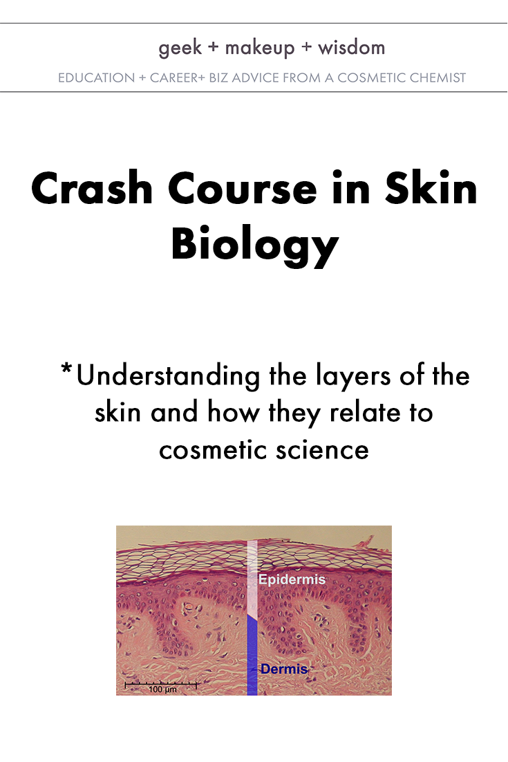
Figure 2. Epidermis and dermis (1)
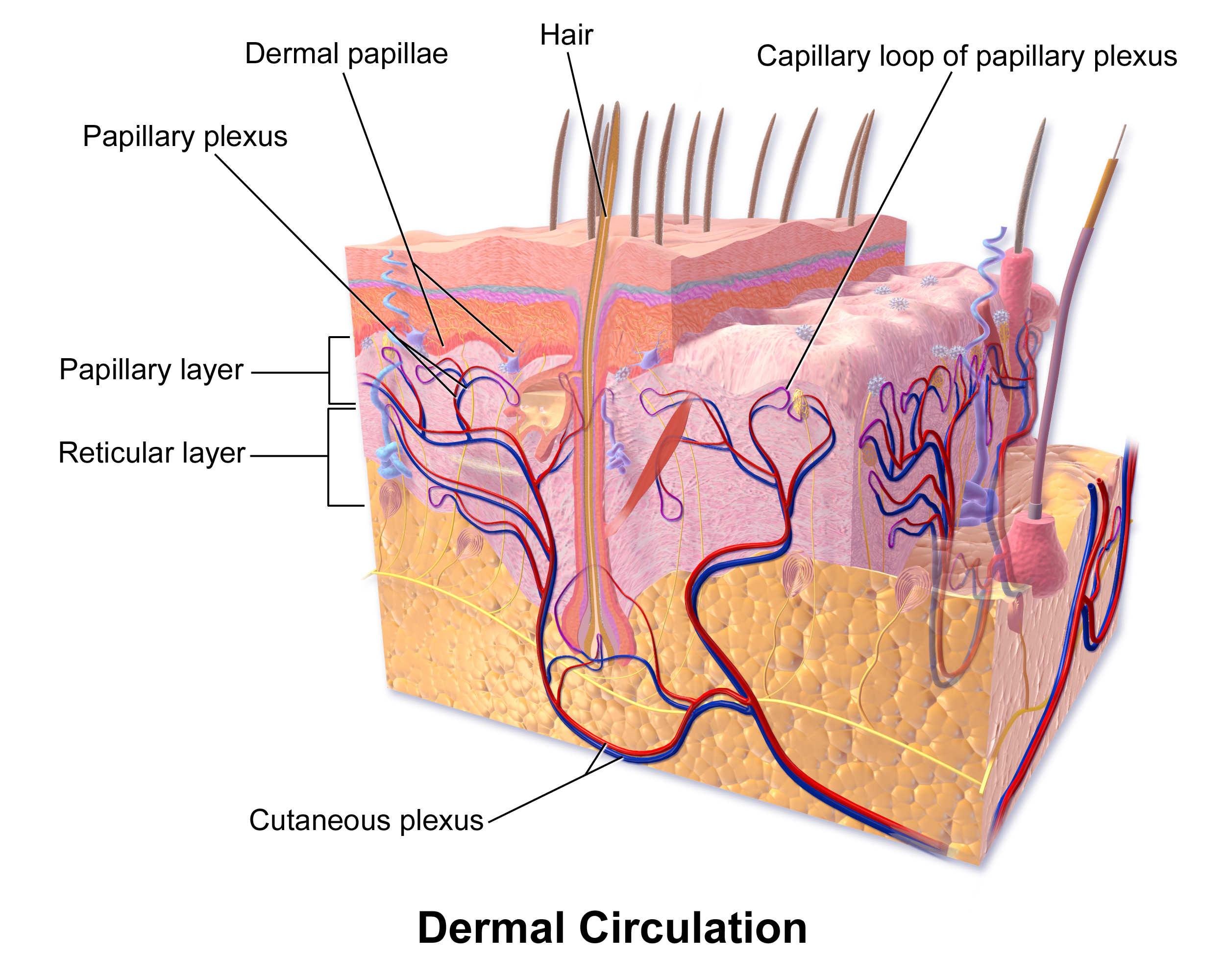
Figure 3. Diagram of skin layers (3)
All right! To summarize:
- Skin cells go through a process called cornification or keratinization, which is its journey through our epidermis as it becomes these keratin-filled cells which protect our bodies.
- We do not absorb water due to the lipid layer that covers our skin.
- In order for products to penetrate our skin, they must disturb the lipid layer.
- If your product acts beyond the upper layer of skin and alters the biology in some way, you have what the FDA defines as a drug, not a cosmetic.
Ok! Hope you enjoyed this post and you learned a lot. Be sure to share it on social media so other budding cosmetic chemists can have a better understanding of skin biology as well. Next week I'll go over the ways ingredients penetrate the skin. If you have any specific questions, let me know in the comments!
References:
1. By Normal_Epidermis_and_Dermis_with_Intradermal_Nevus_10x.JPG: KilbadCropped and labeled by Fama Clamosa (talk) and Mikael Häggström, respectively - Normal_Epidermis_and_Dermis_with_Intradermal_Nevus_10x.JPG (Public Domain)Scale at lower left was created from the an estimation of mean epidermal cell nuclei of 8.6 μm according to the following study:(2011). "Automated identification of epidermal keratinocytes in reflectance confocal microscopy". Journal of Biomedical Optics 16 (3): 030502. DOI:10.1117/1.3552639. ISSN 10833668., Public Domain, https://commons.wikimedia.org/w/index.php?curid=10759481
2. By Mikael Häggström, based on work by Wbensmith - File:WVSOM Meissner's corpuslce.JPG by Wbensmith.Layers were drawn according to image at Home Page of Deborah S. Dempsey, Department of Biological SciencesNorthern Kentucky University > V. SKIN > 2 LAYERS ([1]), CC BY-SA 3.0, https://commons.wikimedia.org/w/index.php?curid=10759398
3. By BruceBlaus. When using this image in external sources it can be cited as:Blausen.com staff. "Blausen gallery 2014". Wikiversity Journal of Medicine. DOI:10.15347/wjm/2014.010. ISSN 20018762. - Own work, CC BY 3.0, https://commons.wikimedia.org/w/index.php?curid=30871443